The Eye Behind the Excellence
There is a moment in every complex dental procedure when the margin between good and exceptional is measured not in millimetres but in micrometres. It is the precise fit of a prosthetic screw. The clean negotiation of a calcified canal. The imperceptible bevel of a veneer margin. These are the moments that define a dentist's career, and they are the moments where technology and training converge.
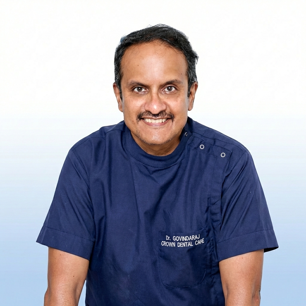
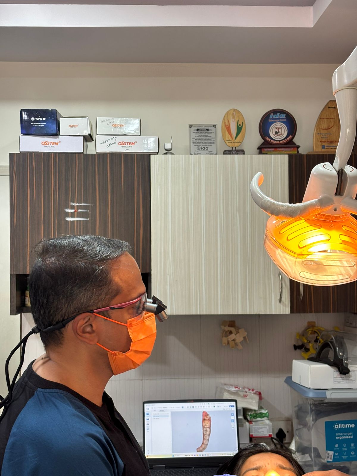
At Crown Dental Care, Dr. Govind has embraced the Ergo Loupe system as a cornerstone of clinical practice — a tool that transforms not just what the clinician sees, but how they think, how they move, and ultimately, how they heal. This blog walks you through four transformative clinical scenarios where enhanced magnification has elevated outcomes from satisfactory to extraordinary.
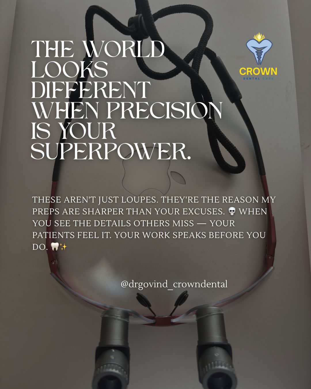
WHY LOUPES?
2.5x magnification. Ergonomic frame engineering. Zero compromise on posture, precision or patient outcomes — the philosophy of Crown Dental Care in one elegant instrument.
Scenario 01 — Implant Prosthetics: The Screw Engagement Advantage
Implant prosthetics represent one of the most technically demanding disciplines in modern dentistry. The restorative phase — and specifically, the engagement of the prosthetic screw — demands a level of precision that is simply unachievable with the naked eye.
The Clinical Challenge
When seating an implant-supported crown, the prosthetic screw must achieve absolute passive fit. Microgaps at the implant-abutment interface of even 10–50 microns can create conditions for bacterial colonisation, peri-implant bone loss, and screw loosening. These failures are often clinically silent until significant damage has occurred. The standard of care is not approximation — it is perfection.
Clinical Scenario: Implant Prosthetic Screw Engagement
Patient presenting with a three-unit implant-supported bridge in the posterior maxilla. Under direct vision, the seating appeared satisfactory. Under 2.5x Ergo magnification, a subtle rotation discrepancy of the abutment was identified prior to final torquing — a finding that would have been invisible without loupes. The screw was re-engaged at the correct angulation, and passive fit was confirmed. Outcome: zero complications at 12-month review.
How Ergo Loupes Transform Implant Prosthetics
- Precision: Real-time visualisation of screw seating confirms complete platform engagement before torquing.
- Detection: Identification of microgaps at the abutment-crown interface during cementation protocols.
- Angulation: Correct angulation of screw driver access channel — eliminating stripping risk on titanium threads.
- Ergonomics: Ergonomic posture preservation during prolonged implant procedures reduces clinician fatigue-induced error.
At Crown Dental Care, every implant prosthetic procedure is performed under magnification as standard. This is not a luxury — it is the protocol that protects both the patient and the restoration.
Scenario 02 — Endodontics: Precision Root Canal Treatment
Root canal treatment remains one of dentistry's most misunderstood procedures — feared by patients, yet when performed with precision, one of the most effective and comfortable interventions available. The quality of endodontic outcomes is inextricably linked to the clinician's ability to navigate anatomy that is, by its very nature, hidden.
The Clinical Challenge
The human root canal system is not a simple tube. It is a complex three-dimensional network of main canals, lateral canals, anastomoses, and apical deltas — variations that remain invisible to the unaided eye. Missed canals are the single most common cause of endodontic failure. In molars, the second mesiobuccal canal (MB2) is missed in up to 45% of cases in non-magnified settings. The consequences — persistent infection, continued pain, eventual extraction — represent a cascade of preventable clinical failure.
Clinical Scenario: Precision Root Canal Treatment — Mandibular Molar
A patient referred from a colleague with persistent post-treatment pain following root canal treatment of a lower right first molar. Periapical radiography revealed no obvious deficiency. Under Ergo magnification during access cavity refinement, a fourth canal — a middle mesial canal — was identified between the standard two mesial canals. This previously untreated canal was the source of the persistent infection. Complete chemo-mechanical preparation and obturation of all four canals resolved the patient's symptoms entirely within one week.
The Loupe Advantage in Endodontics
- Canal Discovery: Identification of extra canals (MB2, middle mesial, palatal accessory) that are routinely missed under direct vision.
- Access Design: Precise straight-line access cavity preparation minimises unnecessary coronal tooth structure removal.
- Calcifications: Detection of canal calcifications and the optimal entry point for negotiation with hand files.
- Wall Inspection: Confirmation of clean canal walls and complete removal of pulp tissue during shaping.
- Crack Detection: Identification of micro-cracks and fracture lines that influence prognosis and treatment planning.
Root canal treatment under Ergo magnification at Crown Dental Care is not merely about treating disease — it is about comprehensively understanding the anatomy before a single instrument is placed. This philosophy has measurably improved first-visit success rates and has eliminated the most common cause of endodontic retreatment: the missed canal.
Scenario 03 — Post and Core: Engineering the Foundation
A post and core restoration is the clinical bridge between a root-treated tooth and its final crown — and like all bridges, its integrity depends entirely on the quality of its foundations. The placement of a post and the fabrication of a core are procedures where magnification moves the outcome from unpredictable to engineered.
The Clinical Challenge
The most critical phase of post placement is canal preparation — specifically, the preservation of adequate gutta-percha apical seal while creating sufficient post space for retention. Perforation of the root, transportation of the canal, and inadequate remaining dentin are among the most serious complications. With root walls that may be as thin as 1.0 to 1.5 mm in the danger zone, the margin for error is vanishingly small. Without magnification, the clinician is operating in a space they cannot fully see.
Clinical Scenario: Post and Core — Maxillary Central Incisor
A patient requiring crown lengthening and post-retained core build-up on a heavily restored maxillary central incisor following trauma and root canal treatment. The root had previously been treated with a silver point (legacy material). Under Ergo magnification, the silver point was visualised and retrieved under controlled conditions using ultrasonic activation. Post space was prepared with precise depth control — confirmed visually — preserving 5mm of apical seal. A fibre post was luted with dual-cure resin composite and the core built to an ideal emergence profile. Crown preparation followed immediately. Outcome: a restoration that has remained stable and asymptomatic for two years at review.
Ergo Loupes in Post and Core Procedures
- Apical Seal: Visual confirmation of gutta-percha level during post space preparation — eliminating the guesswork of radiographic estimation alone.
- Root Anatomy: Detection of root curvatures that influence post selection (prefabricated vs custom cast).
- Wall Assessment: Identification of thin dentinal walls and root concavities that would predispose to perforation.
- Post Fit: Precise fit verification of prefabricated fibre posts before luting — gap detection at the post-canal interface.
- Core Design: Optimised core build-up design with ideal axial wall height, taper and margin placement.
The post and core restoration at Crown Dental Care is approached as an engineering exercise. Every millimetre of tooth structure is considered precious. Every decision — post length, diameter, material, cement — is made with full visual information. Ergo loupes provide precisely that: full information, at the moment it matters most.
Scenario 04 — Veneers: The Art of the Invisible Margin
If implants represent the science of dentistry and endodontics its precision, then veneers represent its art. The porcelain veneer is the finest expression of restorative aesthetics — a wafer of ceramic so thin it must harmonise with tooth structure, light, gingival architecture and the patient's smile line simultaneously. And yet, beneath the artistry lies an unforgiving technical demand: the preparation margin.
The Clinical Challenge
A veneer preparation that is 0.3mm too deep places the margin in enamel-dentine junction territory, risking sensitivity, bond failure and colour instability. A margin that is irregular or unrefined results in a visible line, subgingival inflammation and premature ceramic fracture. The preparation for a porcelain veneer must be feather-light, precisely defined and absolutely uniform — and it must be checked and refined to a standard that no eye unaided by magnification can consistently achieve.
Clinical Scenario: Full Smile Makeover — 10 Porcelain Veneers
A patient presenting with fluorosis staining, mild diastema and lateral incisor peg anomalies requesting full aesthetic rehabilitation with porcelain veneers on upper teeth 13 to 23 (canine to canine). Under Ergo magnification, preparations were made with consistent 0.3–0.5mm enamel reduction using depth-cut burs, with cervical chamfer margins placed precisely at or 0.5mm sub-gingivally in aesthetic zones. Interproximal preparations were extended to break contact without touching adjacent teeth — a manoeuvre visible only under magnification. Temps were fabricated and evaluated over a two-week period. The final veneers, pressed lithium disilicate, were bonded with dual-cure resin cement under magnification — each margin sealed under direct visual confirmation. At one-week review, the patient reported zero sensitivity. At one year, all margins remain invisible clinically and radiographically.
Why Magnification Elevates Veneer Outcomes
- Depth Control: Controlled, uniform preparation depth confirmed in real-time — preventing over-preparation into dentine.
- Margin Refinement: Crisp, defined chamfer margin placement with no rough transitions or chipping of enamel rods.
- Enamel Assessment: Assessment of remaining enamel thickness — the single most important determinant of bond strength.
- Interproximal Precision: Interproximal stripping and preparation without inadvertent contact on adjacent tooth surfaces.
- Seating Confirmation: Cementation under magnification confirms complete marginal seal before light activation of resin cement.
- Cleanup: Post-insertion assessment identifies any residual cement flash — the most common cause of peri-veneer inflammation.
A smile makeover performed at Crown Dental Care is not a compromise between aesthetics and biology. Under Ergo magnification, Dr. Govind achieves both — the invisible margin that reflects no light, the gingival response that shows no inflammation, and the patient who no longer thinks about their smile because they no longer need to.
The Crown Dental Difference: Clinical Advancement as a Standard
The thread that runs through each of these clinical scenarios is not the loupe itself — it is the philosophy that compelled its adoption. At Crown Dental Care, clinical advancement is not an aspiration. It is a standard. A daily commitment to performing every procedure at the highest level currently possible, using every tool that gives the clinician an informational and technical advantage over the unaided eye.
The Ergo Loupe is ergonomically engineered to address one of dentistry's most insidious occupational hazards: musculoskeletal injury. Studies indicate that over 70% of practicing dentists will experience significant neck, shoulder or back pain during their careers — a statistic attributable directly to the hunched, head-dropped posture adopted during close visual inspection without optical aids. With Ergo loupes, the working declination angle is corrected, the clinician works in a neutral spine position, and fatigue — the enemy of precision — is significantly reduced.
CROWN DENTAL STANDARD
Every complex procedure at Crown Dental Care is performed under magnification. Not because it is expected — but because the outcome demands it.
This is not a statement about equipment. It is a statement about values — about the relationship between a clinician and their patient, and the obligation that relationship creates. When a patient sits in the chair at Crown Dental Care, they are not relying on approximation. They are the beneficiary of every advancement Dr. Govind has chosen to adopt, practice and master.
See the Difference. Feel the Difference.
The Instagram post that inspired this blog carried a single headline: See Every Detail. It was not a marketing slogan. It was a clinical declaration.
In the four scenarios described above — implant prosthetic screw engagement, precision endodontics, post and core engineering, and porcelain veneer artistry — the detail that is seen is the difference between a restoration that lasts a lifetime and one that requires revision. Between a root canal that heals and one that fails. Between a smile that the patient forgets about entirely (because it looks perfectly natural) and one they are constantly aware of.
At Crown Dental Care, that detail is never missed. And that is not by accident.
Ready to experience dental care that misses no detail?
Book your appointment at Crown Dental Care